


版权说明:本文档由用户提供并上传,收益归属内容提供方,若内容存在侵权,请进行举报或认领
文档简介
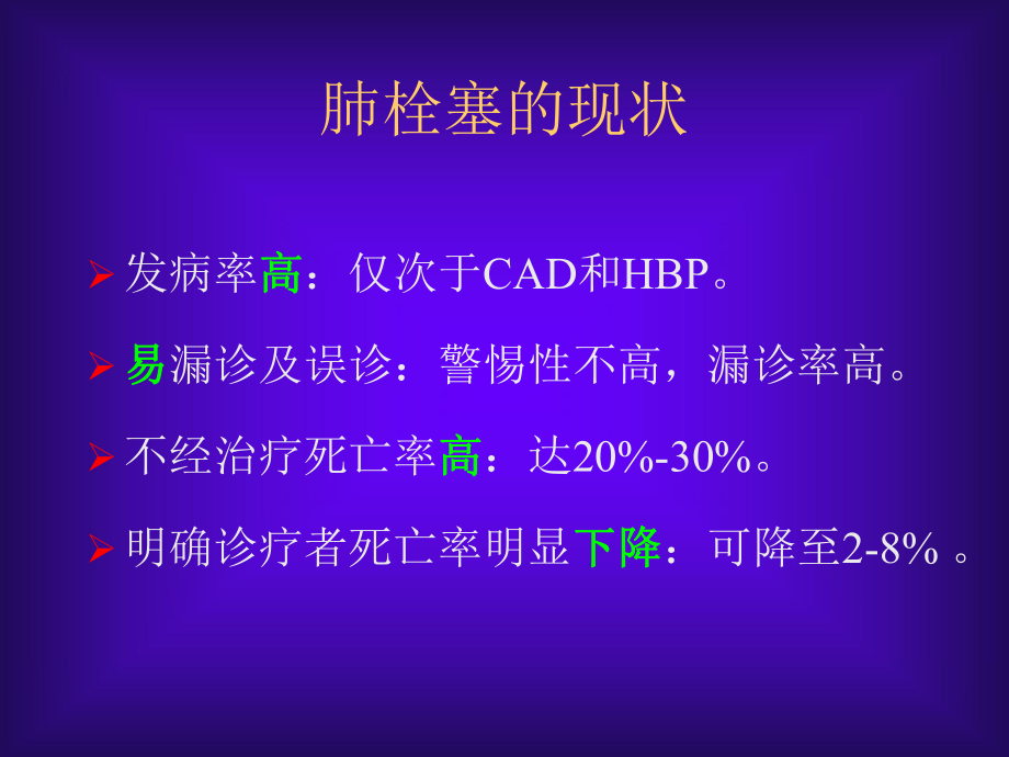
1、肺动脉栓塞的诊治制作XGHRH敬请指正基本概念肺栓塞肺栓塞是以各种栓子阻塞肺动脉系统为其发病原因的一组疾病或临床综合征的总称,包括肺血栓栓塞症,脂肪栓塞综合征,羊水栓塞,空气栓塞等。肺血栓栓塞症肺血栓栓塞症为来自静脉系统或右心的血栓阻塞肺动脉或其分支所致疾病。肺梗死肺梗死为肺动脉发生栓塞后,其支配区的肺组织因血流受阻或中断而发生坏死。肺栓塞的现状发病率高高:仅次于CAD和HBP。易易漏诊及误诊:警惕性不高,漏诊率高。不经治疗死亡率高高:达20%-30%。明确诊疗者死亡率明显下降下降:可降至2-8% 。 EpidemiologyThere is no accurate data for pulm
2、onary embolism because we has limit knowledge of it. In the United States, it is responsible for about 2.3 new cases per 10,000 persons and 50,000 deaths every year.流行病学0 01 12 23 34 46569656970747074757975798084808485898589年龄(岁)年龄(岁)发生率/ 1 0 00患 者 -年发生率/ 1 0 00患 者 -年DVTDVTPEPEArch.Intern.Med.154:86
3、1,1994生存率比较Arch.Intern.Med.154:861,1994随访的年数随访的年数生存的可能性生存的可能性匹配的样本匹配的样本DVTDVTPEPE1.0123Risk Factors for DVT/Pulmonary Embolism (Essential)抗凝血酶缺乏蛋白C缺乏先天性异常纤维蛋白原血症V因子基因突变血栓调节蛋白纤溶酶原缺乏高半胱氨酸血症异常纤溶酶原血症抗心肌碱脂抗体蛋白S缺乏纤溶酶原激活抑制剂过量因子缺乏前凝血酶20210A突变Risk Factors for DVT/Pulmonary Embolism (Second)创伤/骨折外科手术卒中制动高龄恶性肿
4、瘤+化疗中心静脉导管肥胖慢性静脉机能不全心力衰竭吸烟长途旅行妊娠/产后期口服避孕药克隆病、狼疮抗凝剂肾病综合征假体表面粘滞性过高血小板异常深静脉血栓形成原因 分类血流滞缓小腿肌肉静脉丛血栓形成髂股静脉血栓形成静脉壁损伤原发性髂肌静脉血栓形成继发性髂股静脉血栓形成高凝状态股青肿肺血栓与深静脉血栓肺栓塞的大体解剖观肺栓塞的显微镜下观肺栓塞的病理生理肺血管阻塞,神经体液因素或肺动脉压力感受器的作用,引起肺血管阻力增加;肺血管阻塞肺泡死腔气体交换肺泡通气低氧血症V/Q单位气体交换面积二氧化碳刺激性受体反射性兴奋(过度换气)支气管收缩,气道阻力增加肺水肿、肺出血、肺泡表面活性物质减少,肺顺应性降低。肺栓
5、塞后右心功能不全的病生肺栓塞冠状动脉灌注右心室氧需右心室壁张力右心室排血量右心室氧供左心室排血量肺动脉压力右心室后负荷解剖阻塞 神经体液作用右心室扩张/功能不全 右心室缺血室间隔移向左心室低血压体循环灌注左心室前负荷肺栓塞后肺血流动力学变化 前毛细血管高压 血管床减少 支气管收缩 小动脉血管收缩 侧支血管的形成支气管-肺动脉吻合形成 肺内动静脉分流 血流改变: 血流重分布 Westermark征呼吸动力学改变 过度通气: 肺动脉高压 顺应性下降 肺不张 气道阻力增加 : 局限性低碳酸血症 化学介质 临床分型大面积PE(massive PE):休克和低血压;动脉收缩压 3 8.5C体温 3 8.
6、5C喘息喘息Homans征Homans征右室抬举右室抬举胸膜磨擦音胸膜磨擦音第三心音第三心音紫绀紫绀D-二聚体分析检验方法病人数PE发生率% 敏感性 特异性ELISA1579349843快速 ELISA6352410044传统乳胶试验364469255血乳胶试验140259763Adapted from Bounameaux et al, 1997 肺栓塞胸片检查0 01010202030304040505060607070发生率%发生率%正常正常肺不张或实变肺不张或实变胸腔积液胸腔积液胸膜肥厚胸膜肥厚纵隔上抬纵隔上抬肺动脉搏增宽肺动脉搏增宽Westermark征Westermark征心脏增大
7、心脏增大肺水肿肺水肿Peer Review Status: Externally Peer Reviewed by the AMAX-RAY FOR CHESTAtelectasis and parenchymal densities are quite common. The areas of atelectasis are more common in the lower lobe as are the areas of parenchymal densityMost of these densities are caused by pulmonary hemorrhage and ede
8、ma and can be confused with infectious infiltrates or malignant massesPleural effusions are common and most often unilateral despite the fact that most clots are bilateral. These effusions are usually visible when the patient seeks medical attention. They are almost always small, occupying less than
9、 15% of a hemithorax and rarely increase in size after 3 days. Any increase in size after 3 or 4 days should raise the suspicion of a pulmonary infection or re-embolization. Pleural based opacities with convex medial margins are also known as a Hamptons Hump. This may be an indication of lung infarc
10、tion. However, that rate of resolution of these densities is the best way to judge if lung tissue has been infarcted. Areas of pulmonary hemorrhage and edema resolve in a few days to one week. The density caused by an area of infarcted lung will decrease slowly over a few weeks to months and may lea
11、ve a linear scar. A diaphragm may be elevated, reflecting volume loss in the affected lung. The central pulmonary arteries may be prominent either from pulmonary hypertension or the presence of clot in those arteries. Cardiomegally is a non-specific finding but may imply an enlarged right ventricle
12、as seen in the patient who presented with large bilateral pulmonary emboli. A Westermarks sign implies an area of decreased vascularity and perfusion accompanied by an enlarged central pulmonary artery on the affected side. 肺栓塞的心动超声征象直接看到血栓右室扩张右室活动减弱室间隔异常活动三尖瓣反流速度增快肺动脉扩张无吸气性下腔静脉塌陷减弱Br.Heart.J.1994,7
13、2:52室间隔异常活动舒张期收缩期Color-Flow-Doppler-ultrasound非挤压性充盈缺损心电图表现不完全性或完全性右束支传导阻滞、avL的S波1.5mm、avF有Qs波,但无Qs波QRS轴900或不确定肢导联低电压、avF的T波倒置或V1V4T波倒置图12000年8月27日(急诊)ECG大致正常2000年8月29日(门诊)ECG示IRBBB SQTV1V2T波倒置V3V4T波双向Ventilation/Perfusion Lung ScanPIOPED:肺扫描分类与肺动脉造影结果的比较肺扫描肺栓塞肺动脉造影阴性总数有无不肯定高度可疑1021417124中度可疑1052179
14、33364低度可疑391991262312接近正常/正常550274131总计25148024176931J Nucl Med 1993; 34: 1119怀疑PE的患者约25可因肺灌注正常而否定诊断,而且不用抗凝治疗可能是安全的怀疑PE的患者约25具有高度的肺扫描结果,他们可能需要行抗凝治疗其余的患者需要进一步的诊断性检查,而这些检查是更广泛的诊断策略典型肺栓塞 不典型肺栓塞It is high sensitivity but low specificity The differential diagnosis for a ventilation perfusion mismatch inc
15、ludes: acute pulmonary embolus previous pulmonary embolus congenital vascular abnormalities vasculitis, bronchogenic carcinoma, radiation therapy,et al.When a ventilation/perfusion scan does not fit into either the normal or high probability category, then we consider the study to be non-diagnostic
16、and further investigation is required. The majority of cases fall into this category which is characterized by scans with subsegmental defects or defects of any size that match abnormalities on the chest x-ray or the perfusion scan. A low probability category has been suggested by a number of author
17、s. However, as we can see from the PIOPED data this is not a particularly reliable category. Disagreement among experienced readers is common when perfusion defects are small and limit the utility of this category. This study was originally read as showing a small subsegmental defect. Without the ar
18、row, this study has subsequently been called normal by a number of experienced readersConclusionLung scans are sensitive exams that essentially rule out the diagnosis of pulmonary embolus when they are normal. Patients with high probability lungs can often be treated without further workup. Those pa
19、tients with non-diagnostic studies require further diagnostic investigation. CT of Pulmonary EmbolismPulmonary infarcts are more readily identified on CT. Modern CT scanners now have faster acquisition times and are providing a detailed assessment of the lung parenchyma that is not available from th
20、e chest radiograph. The typical appearance of a pulmonary infarct on CT includes a pleural based density with convex borders and a linear strand at the apex of the triangle The apex of the triangle is often truncated and not wedge shaped which corresponds to the normal configuration of a secondary l
21、obule in the lung periphery. Low attenuation areas within the infarct represents viable lung. It is important to note, however, that this appearance is not specific for pulmonary infarction. The differential diagnosis for this abnormality includes infarct, hemorrhage, pneumonia, fibrosis, neoplasia
22、and edemaSince the clinical presentation of pulmonary embolus is usually non-specific, the findings on CT are often the first clinical indication that the patient may be suffering from pulmonary embolus. In addition to visualizing the area of infarction we are often able to see the clot itself. CT h
23、as been show to be especially useful in the assessment of patients with chronic dyspnea and known pulmonary artery hypertension. These patients are often difficult to diagnose as is exemplified by this patient with known sclerodema and pulmonary artery hypertension whose CT unexpectedly showed a lar
24、ge calcified clot in the right pulmonary artery. 肺动脉造影正常肺动脉This selective study was done because of a perfusion defect in the left lower lobe on a ventilation perfusion scan. The first angiographic study was inconclusive. Therefore, a subselective study was done that demonstrated the clot with certa
25、inty. The most reliable signs of pulmonary embolus are: vAn Intraluminal filling defect vAn Abrupt termination of a branch vessel ConclusionAngiography is most accurate in segmental and larger sized arteries. The reproducibility of readings is subsegmental and smaller vessels is poor. Angiography is
26、 a safe procedure that is most accurate when imaging emboli that lodge in segmental or larger arteries. The Diagnosis Algorithm Plasma D-Dimer AssayNormal to Near-NormalLow or Intermediate ProbabilityHigh ProbabilityClinical AssessmentLow ProbabilityIntermediate or High ProbabilityAngiographyPositiv
27、eNegative 500mg/L 500mg/LUltrasonogramNo DVTDVTLung ScanInterpretation CriteriaHigh Probability (80-100% likelihood for PE ):Greater than or equal to 2 large mismatched segmental perfusion defects or the arithmetic equivalent in moderate or large and moderate defects. Intermediate Probability (20-80
28、% likelihood for PE ):1. One moderate to 2 large mismatched perfusion defects or the arithmetic equivalent in moderate or large and moderate defects. 2. Single matched ventilation-perfusion defect with a clear chest radiograph . 3. Difficult to categorize as low or high, or not described as low or h
29、igh. 4. Nonsegmental perfusion defects (e.g., cardiomegaly, enlarged aorta, enlarged hila, elevated diaphragm). 5. Multiple matched V/Q abnormalities, even when relatively extensive, are low probability for PE . The prevalence of PE in patients with extensive matched V/Q defects and no CXR abnormali
30、ty was 14% (low probability). J Nucl Med 1995; 36: 2380-2387Low Probability (0-19% likelihood for PE ) Perfusion defects matched by ventilation abnormality provided that there are: (a) clear chest radiograph and (b) some areas of normal perfusion in the lungs. Extensive matched V/Q abnormalities are
31、 appropriate for low probability, provided that the CXR is clear.Any perfusion defect with a substantially larger chest radiographic abnormality. Any number of small perfusion defects with a normal chest radiograph. J Nucl Med 1995; 36: 2380-2387Diagnostic Criteria for Clinically Suspected Pulmonary
32、 EmbolismPulmonary embolism absentNegative pulmonary angiogranNormal or near-normal lung scanD-dimer level50女女/男比例男比例4:11:1临床经过临床经过进行性恶化进行性恶化稳定一段时间后恶化稳定一段时间后恶化肺灌注扫描肺灌注扫描无节段性灌注缺损无节段性灌注缺损节段性或大片灌注缺损节段性或大片灌注缺损肺动脉收缩压肺动脉收缩压60mmHg60mmHg肺动脉造影肺动脉造影“修剪修剪”征征管腔内充盈缺损管腔内充盈缺损肺动脉造影混淆肺动脉造影混淆的问题的问题血栓血栓“修剪修剪”征也提示征也提示PE确诊确诊肺活检肺活检肺血管镜肺血管镜治疗治疗抗凝;大剂量硝苯地平及静抗凝;大剂量硝苯地平及静注前列环素注前列环素抗凝;抗凝;IVC中断;血栓中断;血栓动脉内膜切除术动脉内膜切除术急性PE的治疗一般处理:送入监护病房,加强生命体征的监护防止栓子脱落,绝对卧床情感支持对症治疗:如咳嗽、发热等急性急性PE呼吸循环支持
温馨提示
- 1. 本站所有资源如无特殊说明,都需要本地电脑安装OFFICE2007和PDF阅读器。图纸软件为CAD,CAXA,PROE,UG,SolidWorks等.压缩文件请下载最新的WinRAR软件解压。
- 2. 本站的文档不包含任何第三方提供的附件图纸等,如果需要附件,请联系上传者。文件的所有权益归上传用户所有。
- 3. 本站RAR压缩包中若带图纸,网页内容里面会有图纸预览,若没有图纸预览就没有图纸。
- 4. 未经权益所有人同意不得将文件中的内容挪作商业或盈利用途。
- 5. 人人文库网仅提供信息存储空间,仅对用户上传内容的表现方式做保护处理,对用户上传分享的文档内容本身不做任何修改或编辑,并不能对任何下载内容负责。
- 6. 下载文件中如有侵权或不适当内容,请与我们联系,我们立即纠正。
- 7. 本站不保证下载资源的准确性、安全性和完整性, 同时也不承担用户因使用这些下载资源对自己和他人造成任何形式的伤害或损失。
最新文档
- 内壁防腐施工合同范本
- 工程扶手采购合同范本
- 商场租赁简单合同范本
- 门面水电开户合同范本
- 地方标准出版合同范本
- 年级组长工作计划范文(5篇)
- 国家开放大学电大《消费者行为学》终结性网考机考题库及答案
- 建筑装饰行业市场行情分析
- 数据处理协议范本
- 企业管理-电工入职笔试题及答案
- 邹平梁邹矿业有限公司矿山地质环境保护与土地复垦方案
- 从目的论看纪录片字幕翻译
- 连锁经营管理理论与实务(全)
- 高考688个高频词汇 word版
- GB/T 9115.4-2000环连接面对焊钢制管法兰
- CAK-13CNC不落轮镟床维修指引
- 项目融资概述课件
- DB225118-2022建筑工程资料管理标准
- 不良资产尽职调查清单
- 小学语文口语交际教学讲座PPT
- 中国电力优质工程奖评审办法
评论
0/150
提交评论